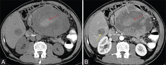
Figure 7 (A and B).

Ileal GIST with hyperdense area within (red arrow in Figure A) and shows no post contrast enhancement (red arrow in B) is hemorrhage within the tumor. Figure B shows heterogeneous enhancement in this tumor with non-enhancing hemorrhagic (red arrow) and necrotic/cystic components within (white arrows). Also there is heterogeneously enhancing liver metastasis (yellow arrow in B)
