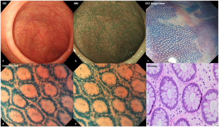
Figure 2.
(a,b) Endoscopic images of mucosal healing (normal vascular and mucosal pattern) with high-definition and narrow-band imaging. (c) Honeycomb appearance of the colonic mucosa with dye chromoendoscopy (methylene blue 0.2%) and Magni view. (d,e) Endocytoscope (methylene blue 0.2%) showed regular crypts and normal spaces between the crypts. (f) Haematoxylin–eosin, original magnification ×200 revealed colonic mucosal healing. (g) Haematoxylin–eosin, original magnification ×400.

