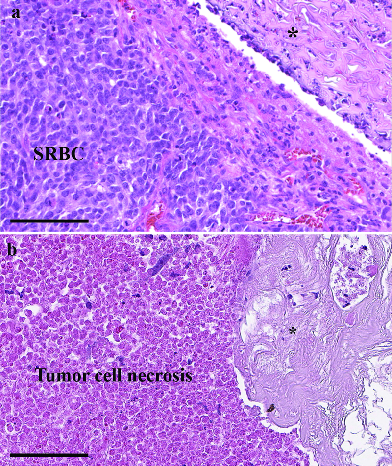
Figure 6.
H&E staining of neuroblastoma xenograft tumor demonstrating (a) viable small round blue cells (SRBC) adjacent to the control silk reservoir (top right corner) and (b) abundant cell necrosis adjacent to silk reservoir loaded with 0.5 mg cisplatin. Asterisk (*) indicates the location of the silk reservoirs. The control and cisplatin-loaded reservoirs show minimal foreign body type reaction, with an absence of infiltrating lymphocytes. Both images are 400 X magnification; scale bar represents 100 μm.

