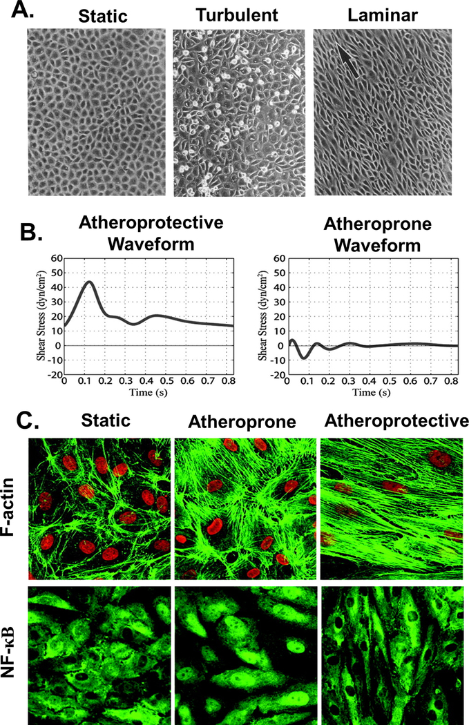
Figure 2. Endothelial monolayer responses to fluid shear stress.
A. Bovine aortic endothelial cells (BAECs) in a confluent monolayer under static conditions (left) exhibit a polygonal configuration with no preferred orientation. ECs exposed to 16 hours of turbulent 0.15 Pa flow (middle) show variable cell shape, lack of alignment, and significant cell rounding. Monolayers after 24 hours of exposure to 0.8 Pa laminar flow (right) show alignment with the direction of flow, indicated by the arrow. Adapted with permission from [60]. B. Representative atheroprotective (left) and atheroprone (right) waveforms were generated from computational simulations of flow patterns in human carotid arteries. C. HUVEC cytoskeletal organization after 24h exposure to static (left), atheroprone (middle), or atheroprotective (right) waveforms generated using a dynamic cone and plate flow system. Atheroprone waveforms enhanced NF-κB nuclear translocation. B&C Adapted with permission from [159]. Copyright 2004 National Academy of Sciences.

