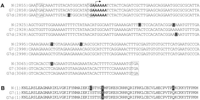
Figure 1.

(A) Partial nucleotide sequences of the P3 cistron of the infectious cDNA clones of Soybean mosaic virus (SMV) strains N (N), G7 and G7d containing the full‐length sequences of pipo. The arrow indicates the starting nucleotide of pipo. The nucleotide sequences within the boxes are pipo in‐frame stop codons upstream of the conserved GA6 motif and the termination codon at the 3′‐end of pipo. The sequences of the GA6 motif at the 5′‐terminus of pipo are shown in italic and boldface. The unique nucleotide of each strain not shared with the others is highlighted. (B) Alignment of the deduced primary amino acid sequences of the PIPO protein of N, G7 and G7d. The unique amino acid of each strain not shared with the others is highlighted. It should be noted that the genomic positions of pipo of G7 and G7d differ from that of N because of the presence of one additional codon upstream of the P3 cistron (GenBank accession numbers AY216010, AY216987 and D00507, respectively).
