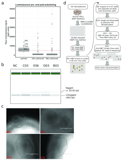
Figure 1. HiBiT-tagging of the membrane surface protein CD46 enables rapid screening for successful CRISPR-mediated knockin in human induced pluripotent stem cells (iPSCs).
a. Boxplots depicting the HiBiT luminescence signal distribution before and after subcloning of CRISPR-targeted cells in the indicated wells. The dashed red line marks the background signal threshold, chosen based on initial standard curve measurements using recombinant HiBiT protein (not shown). b. Capillary electrophoresis following PCR amplification of the targeted CD46 region in non-targeted control (NC) iPSCs and four HiBiT/V5-targeted iPSC populations prior to subcloning. While a clear band-shift can be resolved in targeted cells, the resolution is insufficient to distinguish between a V5 (+42 bp) vs a HiBiT (+32 bp) knock-in. c. Example light micrographs of iPSC colonies following one round of subcloning, revealing healthy colony morphology with clearly defined edges. Scale bar = 1000 µm. d. Summary of the CRISPR/Cas9 HiBiT-based targeting approach. Note that a second round of subcloning may be required depending on the initial targeting efficiency, in line with the original sib-selection approach 4. R, RevitaCell; RNPs, ribonucleoproteins; ssODN, single-stranded oligo DNA.

