
Figure 6.
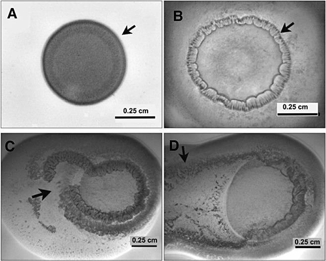
A time lapse series of images showing the different phases on Pnss motility following inoculation on a semi‐solid agar surface. A drop of a 107 cfu/mL suspension was placed on Nutrient agar containing 0.4% agar and 0.4% glucose. (A) A colony of Pnss after 5 h of incubation exhibiting the initiation of colony organization along the periphery (arrow). (B) Periphery of the colony organizing into a palisade ring‐like structure (arrow) after 10 h post‐inoculation. (C) Ring rupture (arrow) after 16 h post inoculation. (D) Rapid surface colonization (arrow) after 30 h. Images used with permission from Herrera et al. (2008).