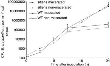
Figure 3.

Survival and multiplication of E. chrysanthemi in sitiens and WT tomato leaf tissue. Leaf discs were taken from infiltrated tissue at the indicated time points, crushed in 50 mm KCl, plated out on LB medium and incubated at 28 °C for 24 h. Colonies of Erwinia chrysanthemi gfp9 strain were identified under UV light by their green fluorescence and counted. Bacterial counts at each time point were compared statistically by the t‐test and no significant differences were detected between sitiens and WT before 24 hpi in three independent experiments. Each data point represents the means + the standard error from one experiment using five or six plants per treatment. At 24 hpi, five macerated and five non‐macerated samples from WT and from sitiens were selected, without representing the frequency of maceration in both genotypes.
