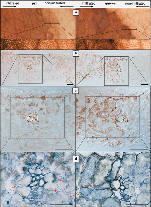
Figure 4.

H2O2 accumulation at the border of E. chrysanthemi lesions in WT and sitiens leaf tissue at 24 hpi. DAB accumulation was evaluated on intact leaf discs (a), and on cross‐sections without (b and c) and with (d) toluidine blue staining. Lesions in WT are characterized by a gradual decrease in chloroplastic H2O2 accumulation towards the outside of the lesion, while sitiens lesions have a clear border between mesophyll cells that contain or lack chloroplastic H2O2. This border in sitiens is located at the site of minor veins and in addition, cells in the vicinity of vascular tissues of the border accumulate extracellular H2O2. Toluidine blue staining (d) reveals bacteria on both sides of the minor vein in WT leaves, while in sitiens no bacteria are present beyond the zone of extracellular H2O2 accumulation. Some bacterial microcolonies are marked with arrows. Scale bar = 50 µm.
