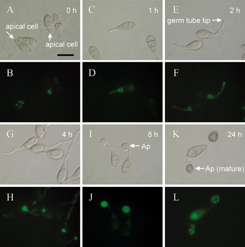
Figure 5.

Localization of a Magnaporthe grisea Chs7p‐GFP fusion protein during infection‐related morphogenesis. A fusion of the M. grisea CHS7 gene with the green fluorescent protein gene (GFP), driven by the CHS7 promoter, was constructed. Localization of Chs7p‐GFP fluorescence within ungerminated conidia was apparent predominantly as large ‘spots’ within the apical cell (A, B), 1 h following germination. Chs7p‐GFP fluorescence was associated with spots within the germ tube and with the conidial cell from which the germ tube arose (C, D). Two hours (E, F) and 4 h (G, H) following germination, Chs7p‐GFP fluorescence was primarily associated with the germ tube apex and/or an apparently vacuolated region of the conidial cell that had given rise to the germ tube (E–H). Nascent appressoria (8 h post‐germination; I, J) showed a strong diffuse Chs7p‐GFP fluorescence, whereas mature appressoria (24 h post‐germination; K, L) showed a punctate distribution of Chs7p‐GFP fluorescence located towards the periphery of the appressorium. Scale bar, 20 µm.
