
Figure 5.
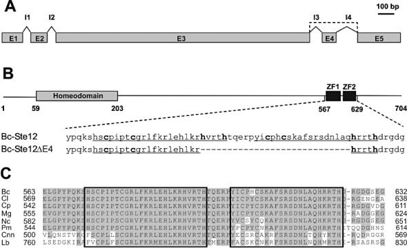
Alternative splicing of Botrytis cinerea ste12 transcripts leads to two different Ste12 proteins. (A) Organization of exons (E1–E5) and introns (I1–I4) in ste12. The broken line indicates the alternative splicing by exon skipping, leading to the shortened ste12ΔE4 transcript. (B) Structures of the predicted Ste12 and Ste12ΔE4 proteins, showing the positions of the homeodomain region and two adjacent zinc finger domains (ZF1 and ZF2, underlined). Alternative splicing results in a shortened protein (Ste12ΔE4) containing only one zinc finger domain. Numbers indicate amino acid positions; conserved cysteine and histidine residues are shown in bold. (C) Alignment of the duplicate zinc finger region in fungal Ste12 proteins. Bc, B. cinerea (FJ374678); Cl, Colletotrichum lindemuthianum (CAD30840); Cnn, Cryptococcus neoformans var. neoformans (AAC01955); Cp, Cryphonectria parasitica (ABE67104); Lb, Laccaria bicolor (XP_001886200); Mg, Magnaporthe grisea (AAL27626); Nc, Neurospora crassa (XP_957811); Pm, Penicillium marneffei (AAK21854).