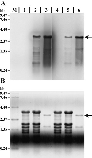
Figure 2.

Evaluation of two 23S rRNA‐specific probes. (A) Northern blot membrane hybridized with DIG‐labelled probe 23S#1 (lanes M, 1–3) or 23S#2 (lanes 4–6). Lanes contain total RNA extracted from uninfected soybean leaf tissue (1, 4; 5 µg), soybean leaf tissue infected with P. syringae pv. glycinea (2, 5; 5 µg), in vitro cultured P. syringae pv. glycinea (3, 6; 0.5 µg) and an RNA size standard (M; 3.0 µg). Hybridization signals specific to the 23S rRNA are indicated by the arrow. (B) Methylene‐blue‐stained membrane prior to Northern hybridization for size estimation, control of total RNA quantity and successful Northern transfer. The same region of the membrane as in A is shown.
