
Figure 4.
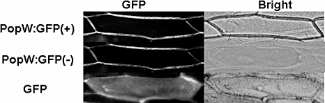
Subcellular localization of PopW in onion epidermal cells. The full‐length popW was transformed into onion epidermal cells via Agrobacterium tumefaciens LBA4404. The localization of fluorescent signals was examined 24 h after transfection under the fluorescence microscope. ‘PopW:GFP’ indicates cells expressing the PopW:GFP fusion protein; ‘GFP’ denotes cells expressing only GFP. (+) and (–) indicate normal cells and plasmolysed cells, respectively. The photographs are fluorescent image (left) and bright field (right).