
Figure 6.
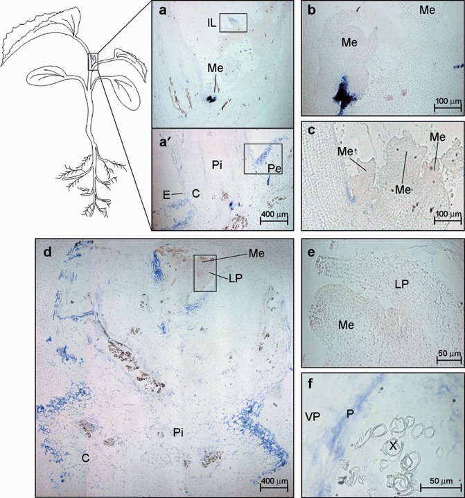
Microscopic imaging of the cellular localization of the Melon necrotic spot virus (MNSV) RNAs by in situ RNA–RNA hybridization with a cvRNA riboprobe in the shoot tip of systemically infected melon plants. The blue colour indicates the presence of target viral RNAs. (a,a′) Longitudinal section of the apical shoot tip showing the cortex (C), the stem pith (Pi), the insertion point of the young leaf petiole (Pe), an immature leaf (IL) and an apical meristem (Me) at 6 dpi. The meristem in panel (a) is enlarged in panel (b). The areas enclosed by both the lower and upper black boxes are enlarged in Fig. 7 in panels (a) and (d), respectively. (c) Longitudinal section of the multi‐meristematic apical shoot tip from a healthy melon plant. (d) Longitudinal section of the apical shoot tip at 10 dpi. Enlarged images of the apical meristem and the leaf primordia (LP) (black box) as well as a vascular bundle showing the MNSV infection into phloem (P) and vascular parenchyma (VP) are displayed on panels (e) and (f), respectively.