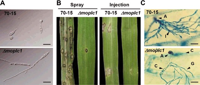
Figure 4.

Appressorium formation and pathogenicity of Δmoplc1. (A) Appressorium formation on a plastic coverslip as an inductive surface was compared between 70‐15 and Δmoplc1. (B) Pathogenicity test. Conidial suspensions were either sprayed onto rice leaves or injected into plant tissues using a syringe for the pathogenicity assay. (C) Penetration defect of Δmoplc1. Ability to penetrate the plant cell was tested by placing the conidial suspension on onion epidermal cells. Scale bar, 20 µm.
