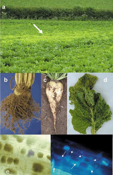
Figure 1.

Symptoms of Beet necrotic yellow vein virus (BNYVV) on sugar beet, and Polymyxa betae, the plasmodiophorid vector of rhizomania. (a) Rhizomania‐infected patch (indicated by arrow) typical of BNYVV infection in the field. (b) Classical ‘root madness’ symptoms in BNYVV‐infected sugar beet. (c) A virus‐free root. (d) Necrotic yellow vein symptom only observed under optimal disease conditions. (e) Long‐lived resting spores in sugar beet roots viewed under the light microscope. (f) Multilobed zoosporangium (solid arrows) inside host root cells after secondary zoospore release. Exit tubes where zoospores were released are visible (dotted arrows). The P. betae zoosporangium had been fixed in situ in 10% formaldehyde, pH 7.2, for at least 3 years prior to being visualised and photographed under the microscope with ultraviolet illumination.
