
Figure 1.
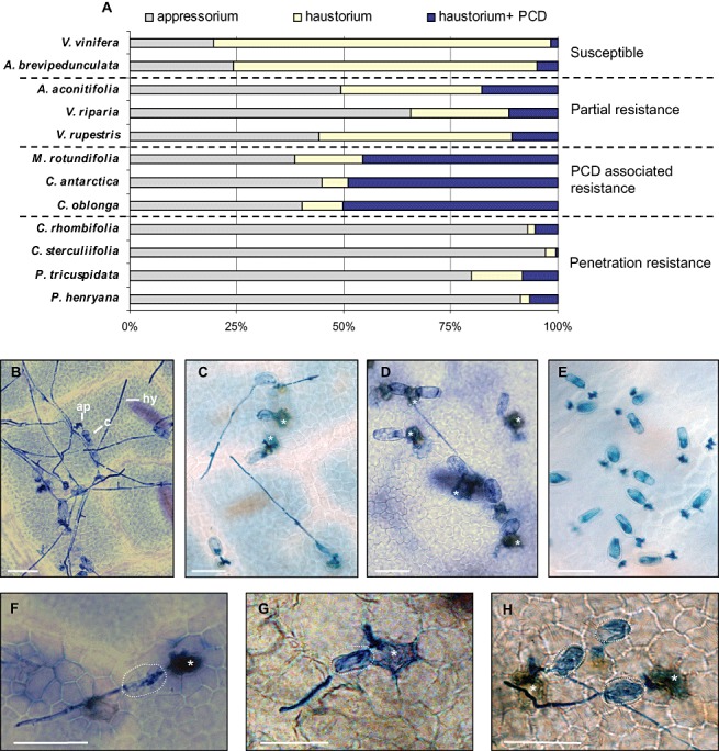
Susceptibility of different Vitaceae species to Erysiphe necator infection. The outcome of E. necator infection on leaves 48 h post‐inoculation. (A) Frequency of E. necator penetration attempts on different Vitaceae members, which concluded in appressorium formation but no penetration, successful penetration and haustorium formation. or a haustorium followed by programmed cell death (PCD) of the penetrated epidermal cell. The frequency of these outcomes in each Vitaceae species leads to susceptibility, partial resistance, PCD‐associated resistance or penetration resistance. Each data point is based on three biological replicates (leaves) on which a minimum of 100 germinated conidia were scored. The data shown are representative of the results obtained in at least two independent experiments. (B–H) Trypan blue staining following E. necator inoculation. (B) Susceptible Vitis vinifera. (C) Partially resistant V. riparia. (D) PCD‐mediated resistance in Muscadinia rotundifolia. (E) Penetration resistance in Parthenocissus tricuspidata. (F) PCD response in M. rotundifolia. (G) PCD response in Cissus antarctica. (H) PCD response in C. oblonga. Asterisks indicate cells which have undergone PCD as stained by trypan blue. Broken white circles indicate the position of a conidium. ap, appressorium; c, conidium; hy, hypha. Scale bars, 50 µm.