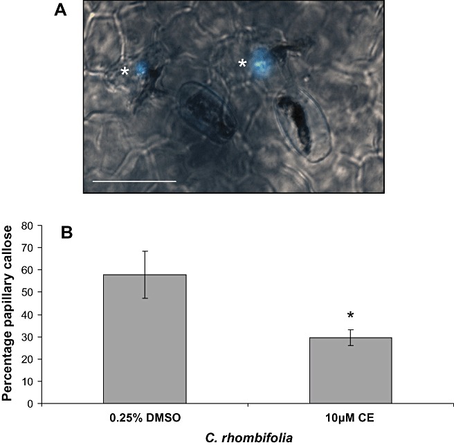
Figure 5.

Erysiphe necator‐induced callose deposition in the papillae of Cissus rhombifolia. (A) Merged bright field and fluorescence image of callose under appressoria marked by an asterisk. (B) Frequency of papillary callose following cytochalasin E or 0.25% dimethylsulphoxide (DMSO) treatment, scored by the presence of callose under spores with an appressorium. Leaves were stained with 0.1% aniline blue. Each data point ± standard deviation is based on three biological replicates (leaves) on which a minimum of 100 germinated conidia were scored. The data shown are representative of the results obtained in two independent experiments. Scale bar, 50 µm. Asterisk indicates a significant difference from DMSO control (P < 0.05; Student's t‐test).
