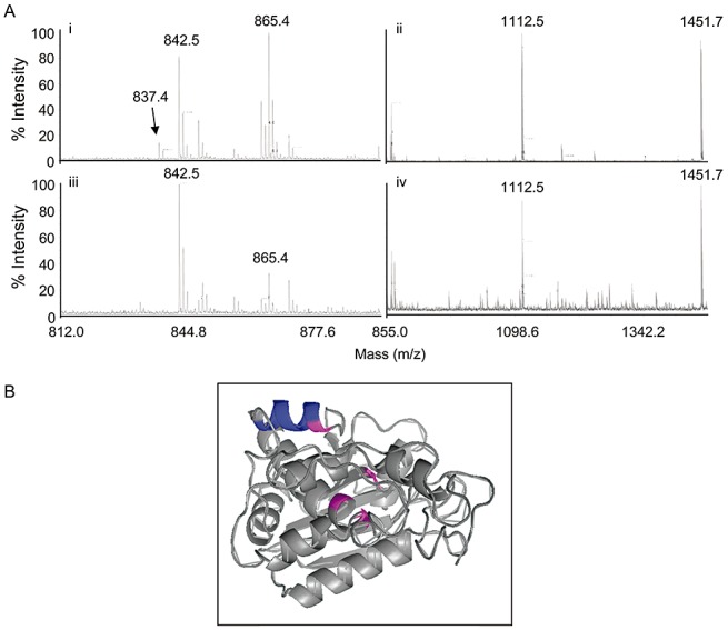
Figure 3.

Mass spectrometric analysis of complexed pokeweed antiviral protein (PAP). (A) Partial peptide spectra of monomeric PAP (i, ii) and complexed PAP (iii, iv) following trypsin digestion. Peak 837.4 corresponds to peptide YPTLESK observed from the digestion of monomeric PAP, but not observed in the spectra of complexed PAP. (B) Molecular model of PAP; β‐strands and α‐helices are indicated, together with the helix containing the heptad YPTLESK (coloured blue). Amino acids essential for enzyme activity are coloured pink.
