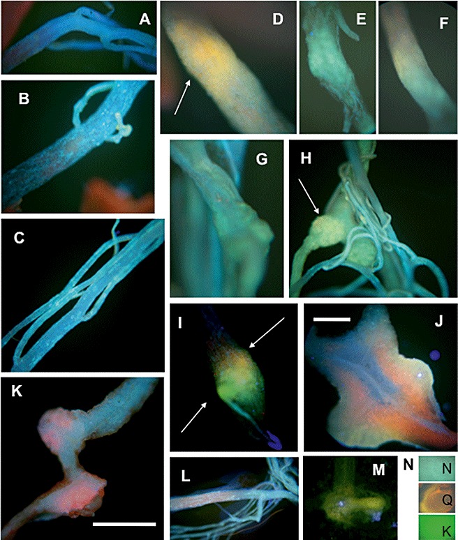
Figure 3.

Accumulation of flavonoids in Arabidopsis control roots and root galls after infection with Plasmodiophora brassicae as shown by staining with 0.5% diphenylboric acid 2‐amino‐ethylester (DPBA) for 2 h. Fluorescence was visualized by excitation with a filter (G365/FT395/LP420 nm). The infected samples were taken at 21 days post‐inoculation (dpi) and control samples were taken at the same age. (A) Control roots of the wild‐type stained with H2O only (the photograph looked the same for infected roots). (B, C) Control roots of the wild‐type showing a blue stain caused by sinapate derivatives. (D–I) Infected roots showing the accumulation of kaempferol (green), naringenin (cyan) and quercetin (orange), taking the colours as preliminary indicators of the respective flavonoid; arrows point to samples for each flavonoid. (J) Cross‐sections showing that flavonoid staining occurs throughout the whole gall and not within single cells. (K) Infected root of the tt4 mutant. (L) Control root of the tt4 mutant. (M) Young gall of the tt7 mutant showing the green coloration typical for kaempferol. (N) Samples of flavonoid standards (K, kaempferol; N, naringenin; Q, quercetin) when incubated directly with the staining solution on a microscopic slide for better comparison. Bar, 0.5 cm (except in J, 0.1 cm).
