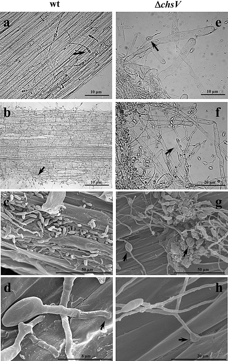
Figure 2.

(a, b, e, f) Light microscopy analysis of tomato roots infected with the wild‐type strain (a, b) or the nonvirulent mutant ΔchsV (e, f). Micrographs were taken 8 h (a, e) or 16 h (b, f) after inoculation. Arrows indicate germlings adhering to the root surface. (c, d, g, h) Scanning electron microscopy analysis of tomato roots infected with the wild‐type strain (c, d) or the nonvirulent mutant ΔchsV (g, h). Micrographs were taken 24 h after inoculation. Arrows indicate penetration sites into the root (d, h) and globular structures in the ΔchsV mutant (g).
