
Figure 4.
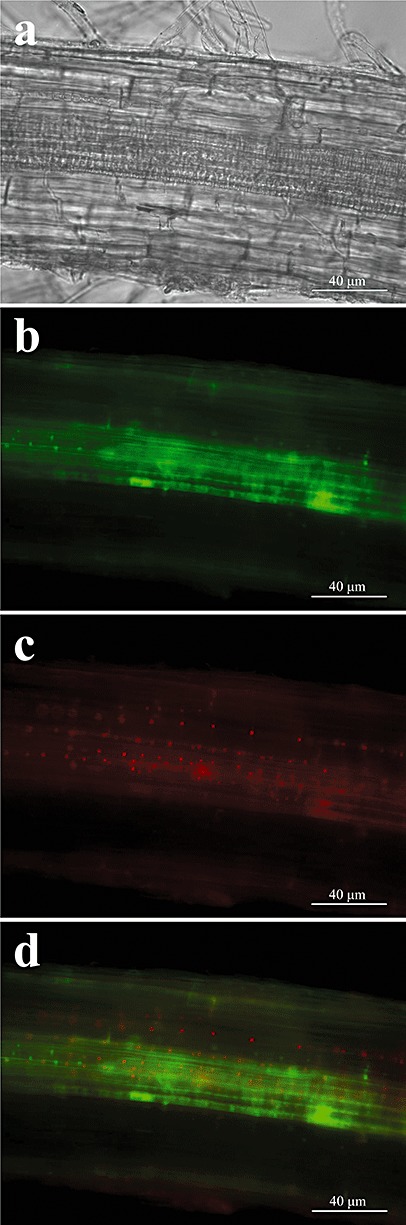
Co‐localization of wild‐type strain [harbouring green fluorescent protein (GFP)‐tagged H1 histone, green] and ΔchsV mutant [harbouring cherry red fluorescent protein (ChFP)‐tagged H1 histone, red] during co‐infection of tomato roots. Differential interference contrast (a) and fluorescence (b, c) micrographs were taken 5 days after co‐inoculation with the wild‐type strain and the ΔchsV mutant in the ratio 5 × 10⁵ : 5 × 10⁶ microconidia/mL, respectively (0.1× : 1×). (d) Images (b) and (c) merged.