
Figure 1.
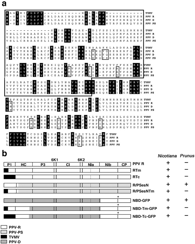
PPV and TVMV P1 protein alignment and PPV/TVMV chimeras. (a) Sequence alignment of PPV and TVMV P1 proteins. Conserved amino acid positions in the sequence alignment are shadowed in black. Open boxes indicate amino acid differences between the PPV isolates. (b) Schematic representation of the different virus constructs used in this work. The pattern assigned to each parental virus is depicted below the constructs. GFP sequences are indicated with an asterisk. The ability of the chimeras to infect Nicotiana species and/or Prunus persicae GF305 is indicated.