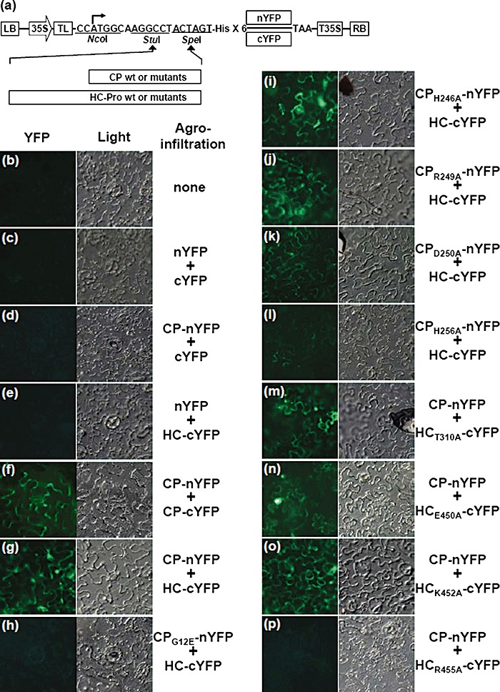
Figure 3.

Detection of in planta interactions between coat protein (CP) and helper component‐proteinase (HC‐Pro) by bimolecular fluorescence complementation (BiFC) assay. (a) Schematic representation of the constructs used for BiFC assay. The basal BiFC vector contains, in sequential order, a left border of T‐DNA (LB), a double 35S promoter (35S), cloning sites, six histidine residues, either nYFP or cYFP sequences, a 35S terminator (T35S) and a right border of T‐DNA (RB). CP and HC‐Pro cistrons and their mutants were in‐frame inserted into the basal BiFC vectors utilizing StuI and SpeI sites. The constructed clones were transferred into Nicotiana benthamiana leaves by agroinfiltration in pairwise combinations as follows: none (healthy; b), nYFP + cYFP (c), CP‐nYFP + cYFP (d), nYFP + HC‐cYFP (e), CP‐nYFP + CP‐cYFP (f), CP‐nYFP + HC‐cYFP (g), CPG12E‐nYFP + HC‐cYFP (h), CPH246A‐nYFP + HC‐cYFP (i), CPR249A‐nYFP + HC‐cYFP (j), CPD250A‐nYFP + HC‐cYFP (k), CPH256A‐nYFP + HC‐cYFP (l), CP‐nYFP + HCT310A‐cYFP (m), CP‐nYFP + HCE450A‐cYFP (n), CP‐nYFP + HCK452A‐cYFP (o) and CP‐nYFP + HCR455A‐cYFP (p). The reconstructed yellow fluorescent protein (YFP) signals were observed in the epidermal cells using epifluorescence microscopy at 3 days post‐infiltration (dpi). Co‐expression of (g), (i), (j), (k), (m), (n) and (o) induced fluorescence signals. Co‐expression of (l) induced weak but detectable fluorescence. YFP signals from the co‐expression of (f) were used as a positive control. Images for fluorescence emitted by YFP (left) and for the transmitted light mode (right) are shown.
