
Figure 2.
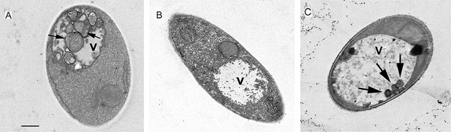
Starvation‐induced autophagosome accumulation in wild‐type, Δatg8 and Δatg1 Ustilago maydis cells. Wild‐type and Δatg8 cells were incubated for 5 h in medium lacking a carbon source (MM – C). Transmission electron images show the accumulation of autophagic bodies (arrows) within the vacuole (v) of wild‐type cells (A). Note the absence of these structures from vacuoles of Δatg8 cells (B). Smaller vesicles occasionally appeared in vacuoles of Δatg1 (C). At least 40 cells were inspected per sample. Scale bar, 1 µm.