
Figure 5.
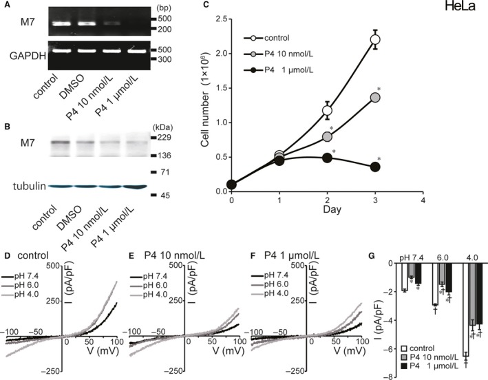
Effects of progesterone (P4) treatment on molecular and functional expression of TRPM7 and cell proliferation in HeLa cell. (A) Detection of TRPM7 and GAPDH mRNAs by RT‐PCR. Treatment with progesterone (10 nmol/L or 1 µmol/L) for 72 h suppressed TRPM7 mRNA expression (compared to DMSO‐treated or untreated control) without affecting GAPDH mRNA expression. Data are representative of duplicate experiments. (B) Immunoblots of the protein extracts of HeLa cells for TRPM7 protein. Progesterone treatment reduced expression of TRPM7 protein. Data are representative of triplicate experiments. (C) Effects of progesterone on cell proliferation evaluated at 1, 2, and 3 days after culture. Gray and filled circles represent the number of HeLa cells treated with 10 nmol/L or 1 µmol/L progesterone, respectively. Open circles represent the number of control cells. (D, E, F) Representative I–V relationships of cationic currents under ramp clamp from −100 to +100 mV in untreated control and progesterone‐treated cells. (G) Summarized data showing the whole‐cell current densities recorded at −100 mV in untreated control cells (white columns), 10 nmol/L (gray columns) and 1 µmol/L progesterone‐treated cells (black columns). Acid treatment (pH 6.0 or 4.0) significantly reduced whole‐cell currents compared to untreated control cells (n = 7–15). Each column represents the mean ± SEM (vertical bar). *Significantly different (P < 0.05) from the control values. †Significantly different (P < 0.05) from the values at pH 7.4.