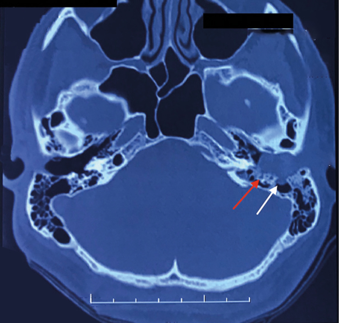
Figure 1.

High resolution computed tomography of temporal bone. Axial section at external auditory canal (EAC) level showing impacted soft tissue density with erosion of posterior the EAC wall (white arrow) and erosion of the anterior wall of the vertical facial canal (red arrow)
