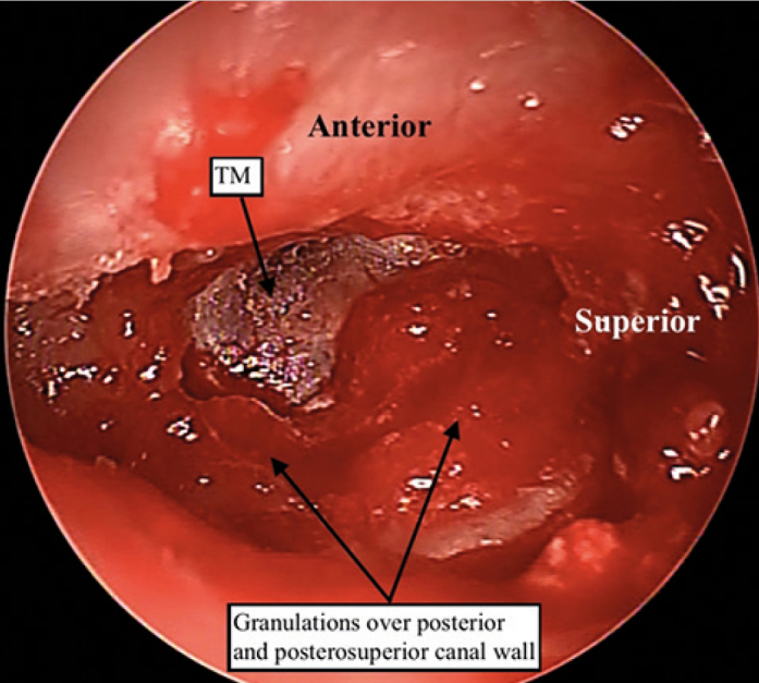
Figure 3.

Left ear intraoperative pictures showing the expanded medial external auditory canal (EAC) with extensive granulations involving the posterior and the inferior part of the EAC encroaching onto the posterosuperior tympanic membrane. (Magnification under microscope X6.4)
