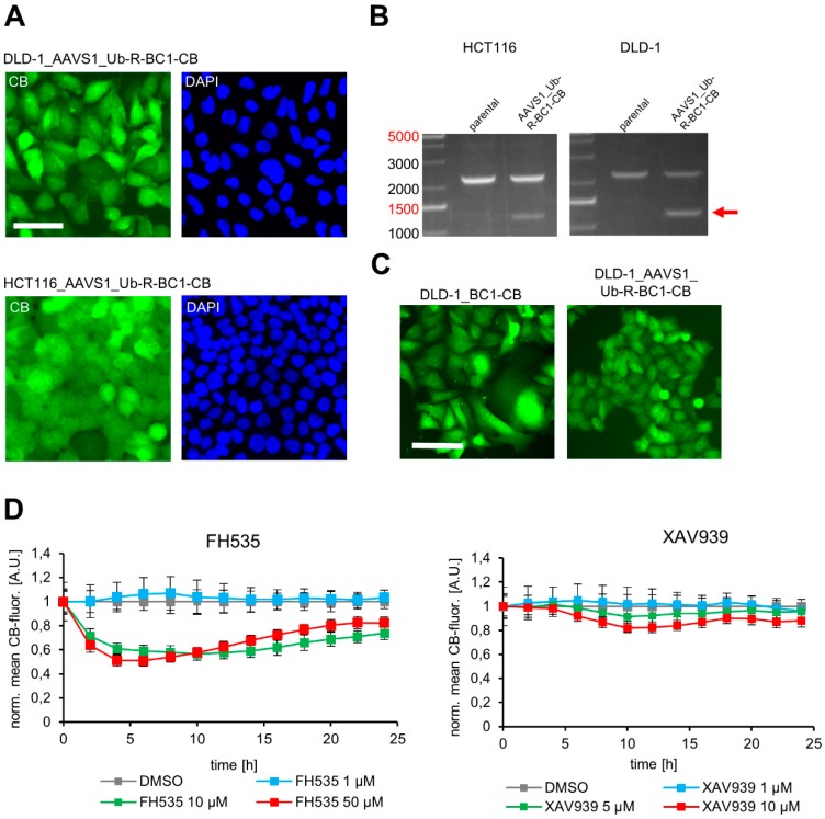
Figure 6.
Generation of colorectal carcinoma cell lines with stable AAVS1 integration of the turnover-accelerated Ub-R-BC1-CB encoding transgene. (A) Representative fluorescence images of isolated DLD-1 (upper panel) and HCT116 (lower panel) clones stably expressing Ub-R-BC1-CB. Nuclei were stained with DAPI, scale bar: 50 µm. (B) Genotyping of HCT116_AAVS1_Ub-R-BC1-CB and DLD-1_AAVS1_Ub-R-BC1-CB. Genomic DNA of host cells was extracted and subjected to PCR using the primer strategy illustrated in Figure 4B. Corresponding PCR products are indicated by arrow. (C) Representative fluorescence images of DLD-1 cell lines stably expressing the indicated BC1-CB. Left image illustrates DLD-1_BC1-CB cells generated by random integration of the non-modified BC1-CB. The right image shows DLD-1_AAVS1_Ub-R-BC1-CB cells generated by CRISPR/Cas9-mediated site-directed integration of the ubiquitin fused BC1-CB, scale bar: 50 µm. (D) Quantification of nuclear CB fluorescence in DLD-1_AAVS1_Ub-R-BC1-CB cells upon compound treatment. DLD-1_AAVS1_Ub-R-BC1-CB were either treated with the indicated concentrations of FH535 or XAV939 or treated with 0.01% DMSO as control. Cells were continuously imaged every 2 h for up to 24 h. Fluorescence intensity of nuclear CB-signal was quantified and the fluorescence values were normalized to the DMSO control and plotted against time (n = 2, >200 cells each). Error bars: S.D.

