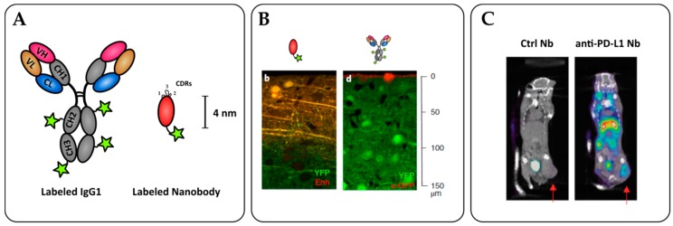
Figure 3.
Nanobodies as potent tools for tumor immunoimaging. (A) Nanobody labelling strategies allow for site-specific and oriented conjugation. (B) Penetration of an anti-GFP nanobody (left) or full-size IgG (right) within an YFP-expressing brain tissue in vitro. Adapted from Fang T. et al. [120]. (C) SPECT/CT imaging of PD-L1 positive mouse lung epithelial cell line TC-1 in C57/BL6 mice with radiolabeled 99mTc nanobodies 1 h after injection. The arrows indicate the tumor site. Adapted from Broos K. et al. [124].

