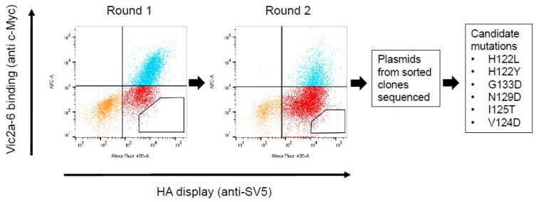
Figure 3.
Cell sorting and isolation of candidate mutations interfering with the Vic2a-6 sdAb binding. Flow cytometry plots for two rounds of negative sorting for loss of binding to Vic2a-6, by gating cells in the lower right quadrant of the FACS dot plot, as indicated. Plasmids from the sorted yeast clones were sequenced and mutations in the HA which interfered with sdAb binding to HA, were identified. Residue numbering was according to B/HongKong/8/73 [29].

