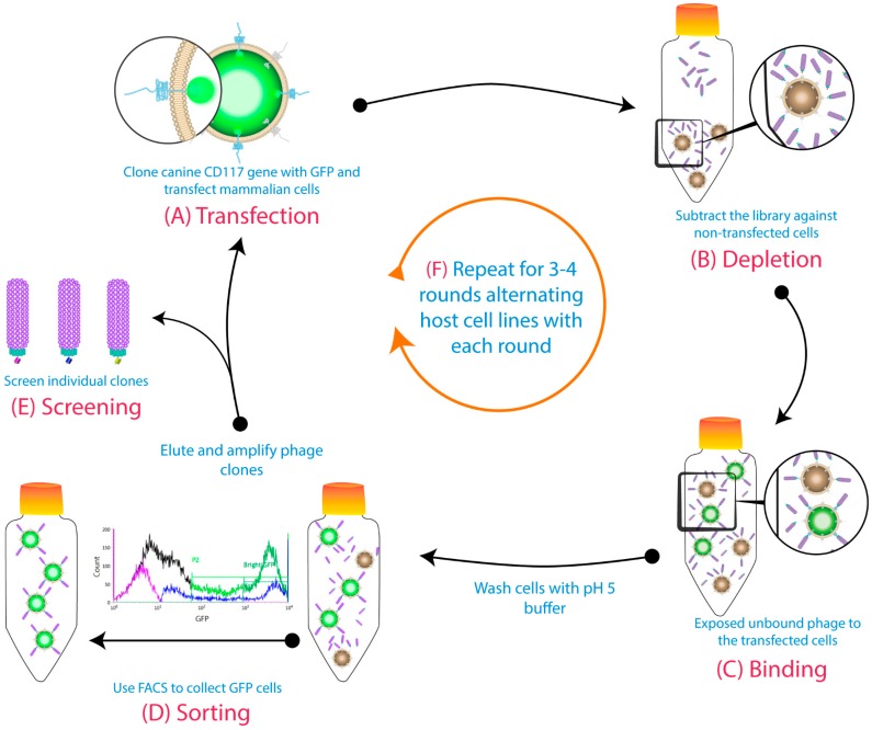
Figure 1.
Cell-based biopanning protocol using cell sorting and alternative host cell lines: (A) The gene for the target membrane protein was cloned in-frame with Green Fluorescent Protein (GFP) to be expressed on the cell surface. (B) The phage library was subtracted on untransfected cells. (C) Then, the free phages were panned against transiently transfected cells. (D) A pH 5 wash step was conducted before GFP-positive cells were collected using fluorescence-activated cell sorting (FACS). The binders from the sorted cells were eluted with a low pH buffer, then amplified for the subsequent rounds. (E) Individual clones were screened at the end of biopanning campaigns. (F) The host cell line was alternated each round (first round: Chinese hamster ovary (CHO) cells; second round: Human embryonic kidney 293 cells (HEK293), etc.). Figure shows methodology described by Jones et al. (2016) [18].

