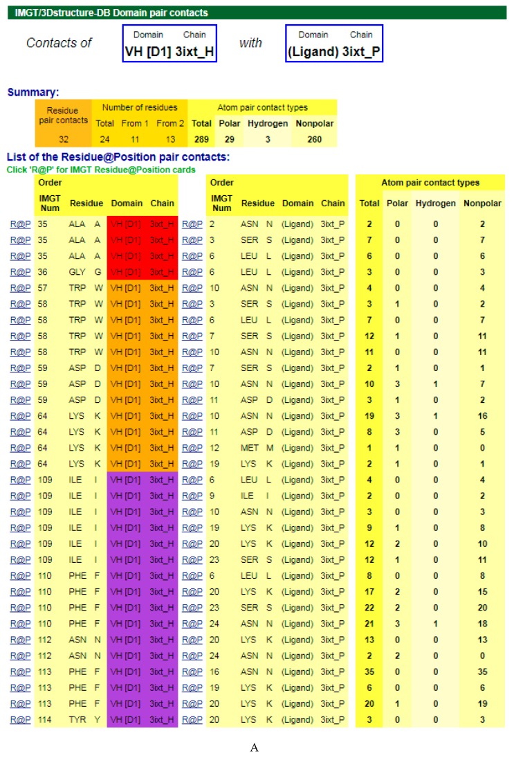
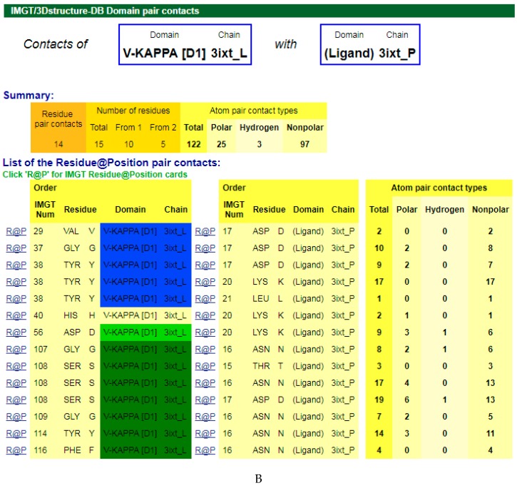
Figure 3.
V-DOMAIN Contact analysis results. Reproduced with permission from IMGT®, the international ImMunoGeneTics information system®, http://www.imgt.org. (A) IMGT/3Dstructure-DB domain pair contacts between the VH domain of motavizumab (3ixt_H) and the Fusion glycoprotein F1 (ligand) (3ixt_P). (B) IMGT/3Dstructure-DB Domain pair contacts between the V-KAPPA domain of motavizumab (3ixt_L) and the Fusion glycoprotein F1 (ligand) (3ixt_P). ‘Polar’, ‘Hydrogen bonds’, and ‘Nonpolar’ were selected prior to display, in ‘Atom contact types’. Amino acids belonging to the CDR1-IMGT, CDR2-IMGT and CDR3-IMGT are colored according to the IMGT color menu (red, orange, and purple, respectively, for VH; blue, light green and green, respectively, for V-KAPPA). In this 3D structure, all but one of the amino acids contacting the antigen belong to the CDR-IMGT. Clicking on R@P gives access to the IMGT Residue@Position cards [9,10,11].


