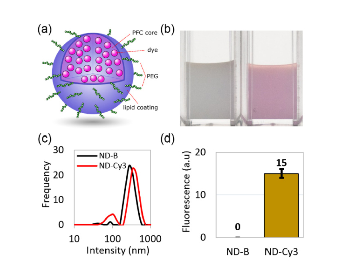
Fig. 1.
(a) Schematic of a nanodroplet with lipid coating and PFC core containing dye molecules (ND-Cy3); (b) a photograph of washed preparations of “blank” nanodroplets with no dye (ND-B, left) and ND-Cy3 (right); (c) size distributions of nanodroplets measured by dynamic light scattering (intensity distribution); (d) difference in fluorescence between washed ND-B and ND-Cy3 nanodroplets at the same concentrations.

