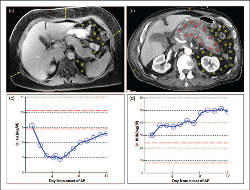
FIGURE 1.
Abdominal imaging showing how location of fat influences the severity of pancreatitis. (a) MRI of a 50-year-old female (weight 85 kg, BMI 34.4) on the second day of biliary pancreatitis showing predominantly subcutaneous fat (yellow arrows) compared with visceral fat (area with yellow dots) with no fat necrosis. The patient had a mild course and was symptom free after 3 days of conservative management. (b) CT scan of a 83-year-old male (weight 84 kg, BMI 26.6) 2 weeks into alcoholic pancreatitis showing little subcutaneous fat (yellow arrows), but a large amount of visceral fat (dots in the peritoneal cavity). The fat around the pancreas was involved in peripancreatic fat necrosis (red dots), whereas the more distant visceral fat remained uninvolved (yellow dots). The clinical course was associated with early hypocalcemia (c), renal failure of >48 h (d) and the patient requiring ventilator support. AP, acute pancreatitis; CT, computerized tomography.

