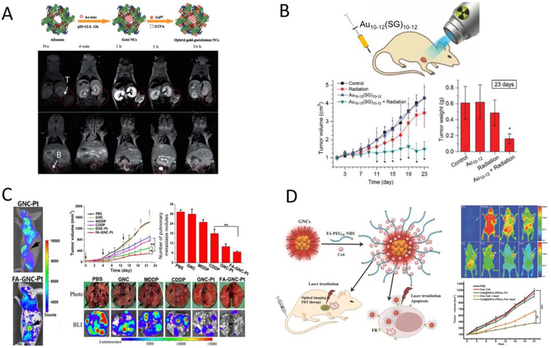
Fig.8.
(A) Schematic of the synthetic route to the hybrid gold-gadolinium nanoclusters (top); in vivo MRI images of MCF-7 tumor-bearing mice (tumor and bladder were indicated) at multiple time points after IV injection of the hybrid gold-gadolinium nanoclusters (bottom). (B) Illustration of ultrasmall Au10–12(SG)10–12 nanoclusters as cancer radiosensitizers (top); time-course studies of the tumor volumes and tumor weights (at 23 days post injection) for different experimental groups after radiation therapy (bottom). (C) In vivo fluorescence imaging of the cisplatin prodrug conjugated fluorescent gold nanocluster (GNC-Pt) and GNC-Pt linked with folic acid (FA-GNC-Pt) in 4T1 tumor-bearing mice (left); anti-cancer efficacy of FA-GNC-Pt and other control studies on the 4T1 primary tumor volume as well as the number of lung metastasis nodules examined by bright field and bioluminescence imaging (right). (D) Schematic illustration of the preparation of the folic acid-conjugated photosensitizer (Ce6)-loaded fluorescent AuNCs (Ce6@GNCs-PEG-FA) and their applications in targeted imaging and photodynamic therapy (left); in vivo fluorescence imaging and tumor growth trend after photodynamic therapy using Ce6@GNCs-PEG-FA (right). (Adapted or reprinted with permissions from Royal Society of Chemistry for (A) Ref. [290], John Wiley and Sons for (B) Ref. [67] and (D) Ref. [281], Ivyspring International Publisher for (C) Ref. [296].)

