FIGURE 1.

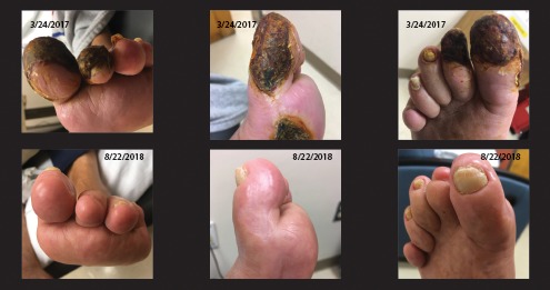
Evolution of wound appearance in a patient at baseline (top) and infusion 48 (bottom). Last revascularization procedure was 3 months before top row of images were taken, resulting in progressive deterioration.

Evolution of wound appearance in a patient at baseline (top) and infusion 48 (bottom). Last revascularization procedure was 3 months before top row of images were taken, resulting in progressive deterioration.