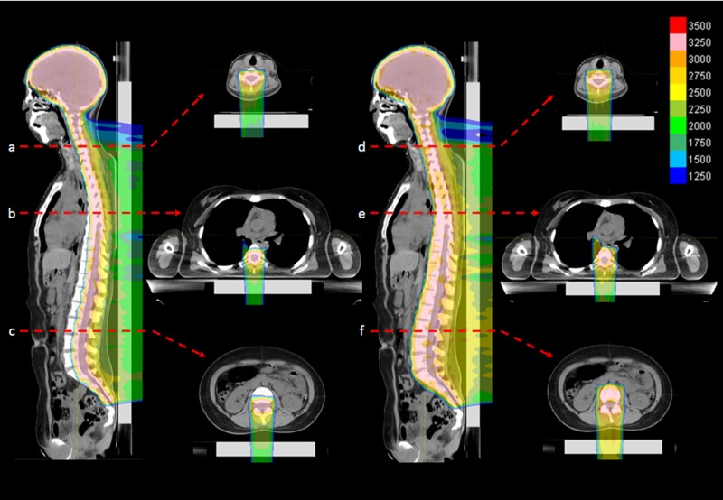
Figure 1.
(Left) An example of actual dose distribution of VBSipCSI in an adolescent patient (Case no. 3 in Table 1): cervical spine (top), thoracic spine (middle) and lumbar spine (bottom) levels. (Right) Simulated dose distribution of ipCSI without VBS for the same patient, which was not used for the patient.

