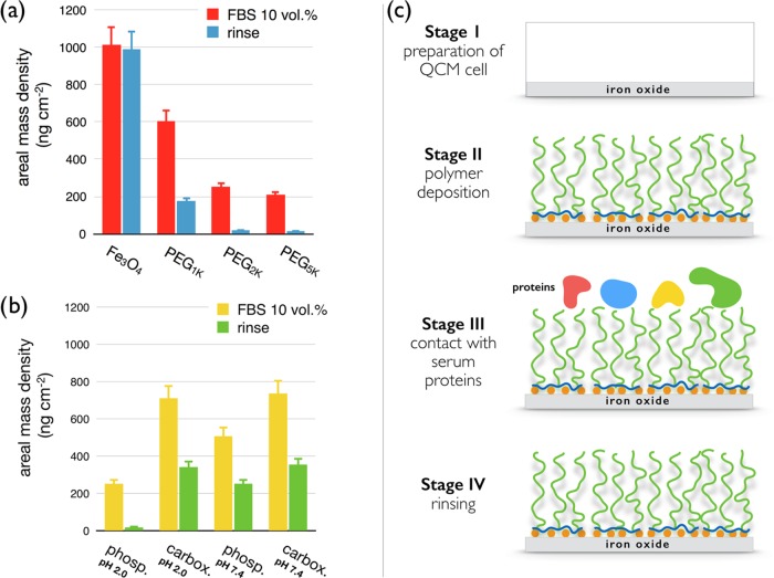
Figure 8.
(a) Protein areal mass densities on PEGylated iron oxide substrates for different PEG molecular weights. (b) Same as that in (a) for different brush formation conditions. With a protein resistance of more than 99%, phosphonic acid PEG5K copolymers deposited at pH 2.0 is the most repellent layer. (c) Illustration of the different adsorption and rinsing stages used in the QCM-D protocols, including cell preparation (stage I), polymer deposition (stage II), exposition to serum proteins (stage III), and final rinsing (stage IV).

