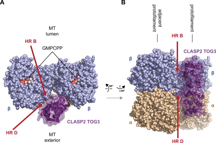
Fig 7. M.m. CLASP2 TOG3 is predicted to engage laterally associated tubulin on the microtubule lattice.
(A) Model of M.m. CLASP2 TOG3 superpositioned on a microtubule. Shown are two laterally-associated tubulin heterodimers from neighboring protofilaments. M.m. CLASP2 TOG3 is shown in dark purple, modeled bound to the tubulin heterodimer shown at right (akin to the single-tubulin heterodimer binding mode depicted in Fig 6A). The model generated of M.m. CLASP2 TOG3 bound to free tubulin (Fig 6A) was superpositioned onto the lattice coordinates of GMPCPP-bound tubulin (PDB accession code 3JAT [53]). The tops of the β-tubulin subunits are shown (plus end oriented towards the viewer), looking into the bore of the microtubule with the luminal region oriented above and the microtubule exterior oriented below. (B) Model as shown in (A), viewed from the microtubule exterior with the plus end oriented up. β-tubulin is shown in lavender, α-tubulin is shown in wheat. Potential M.m. CLASP2 TOG3 HR B and HR D contacts with the laterally-associated tubulin subunit are demarcated with red arrows.

