Figure 2.

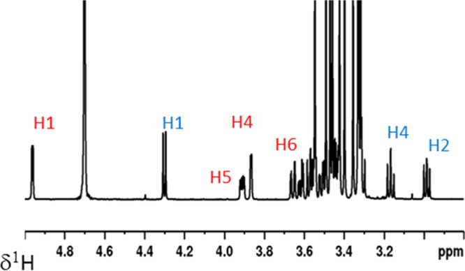
One-dimensional 1H NMR spectrum of an equimolar mixture of β-pMeGlc(2)/α-pMeGal(3) in D2O at 298 K (ca. 25 mM, 600 MHz). Assignments of the signals of β-pMeGlc(2) and α-pMeGal(3) are highlighted with blue (2) and red (3).

One-dimensional 1H NMR spectrum of an equimolar mixture of β-pMeGlc(2)/α-pMeGal(3) in D2O at 298 K (ca. 25 mM, 600 MHz). Assignments of the signals of β-pMeGlc(2) and α-pMeGal(3) are highlighted with blue (2) and red (3).