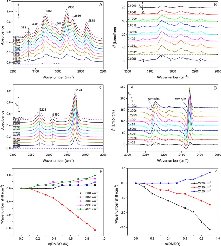
Figure 3.
ATR–FTIR (A) and excess IR (B), indicating the spectra of C–H stretching vibrations in [Bpy][DCA]/DMSO-d6 mixtures from 3200 to 2800 cm–1, and ATR–FTIR (C) and excess IR (D), indicating the spectra of C≡N in [Bpy][DCA]/DMSO mixtures from 2300 to 2050 cm–1. E and F showing the wavenumber shifts of C–H and C≡N vibrations during the dilution, respectively.

