
Figure 1.
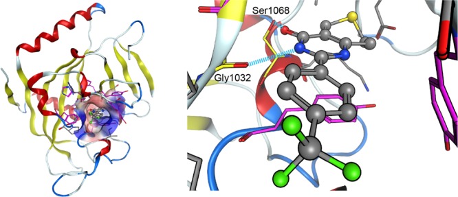
Tankyrase 2 inhibitor (C01) in the nicotinamide binding pocket. The left-hand figure shows the structure of the tankyrase 2-inhibitor (C01) complex (PDB entry: 3KR8). The molecular surface representation shows the nicotinamide binding pocket that accommodates the inhibitor. Here, the inhibitor is shown by a ball and stick model. The hydrophobic and hydrophilic regions in the binding pocket are drawn in blue and red, respectively. The amino acid residues constituting the binding pocket are shown by stick models. Among these amino acid residues, aromatic amino acid residues are drawn in purple. The right-hand figure shows the inside of the binding pocket of the left-hand figure. Gly1032 and Ser1068 in the tankyrase 2 protein form hydrogen bonds with the inhibitor. It is expected that Gly1032 and Ser1068 would form similar hydrogen bonds with other inhibitors.