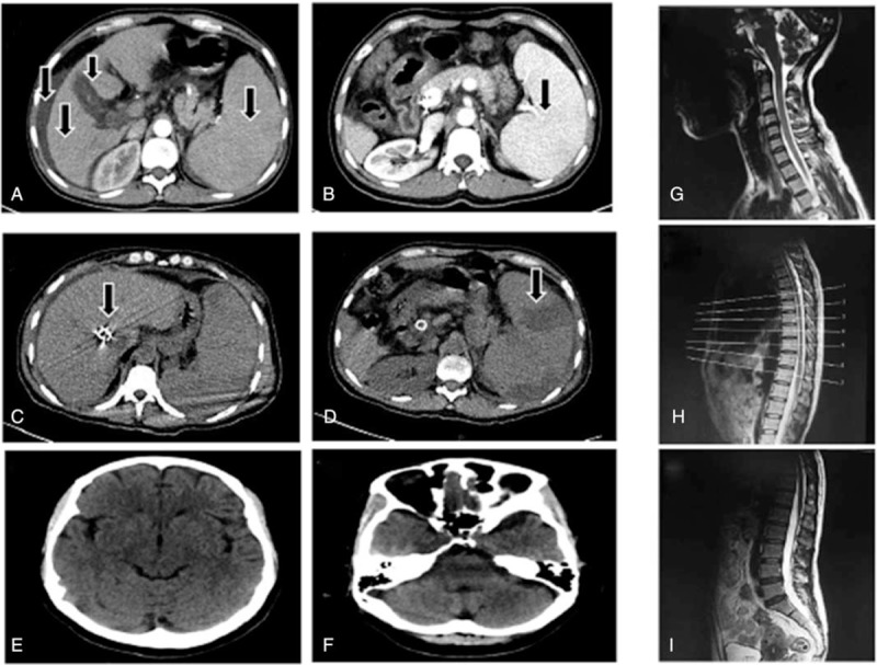
Figure 1.

Abdominal CT image of the patient. Arterial phase (A) and venous phase (B) of CT abdomen showing liver cirrhosis, splenomegaly, ascites, cholecystitis. And CT scan (C, D) showing implantation of stent and infarctions of spleen after portacaval shunts. Cranial CT (E, F) indicated the possibility of empty sella, no other abnormality. Sagittal T2-weighted MR images of the cervical (G), thoracic (H), and lumbar (I) spine, there was no pressure on the spinal cord. CT = computed tomography, MR = magnetic resonance.
