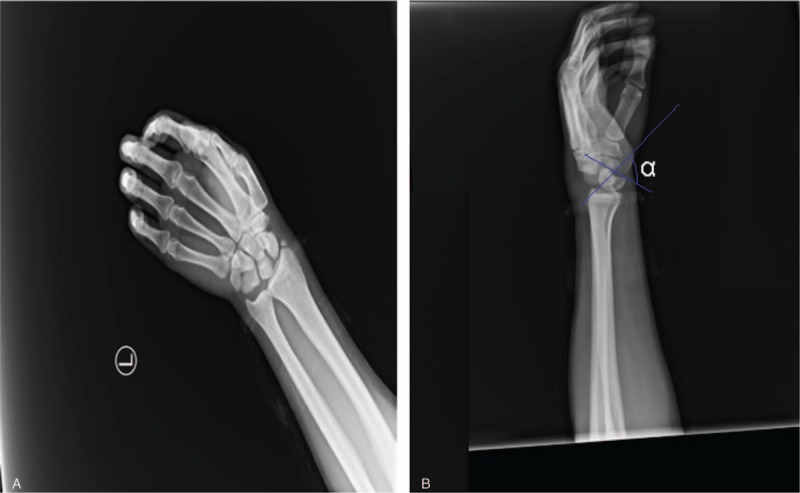
Figure 1.

A, The initial x-ray image after injury showing lunate dislocation and fracture of the styloid process of the radius. B, Lateral view of the left wrist showing volar dislocation of the lunate (arrow) and the “spilled teacup” sign. The scapholunate angle (α) is >70°.
