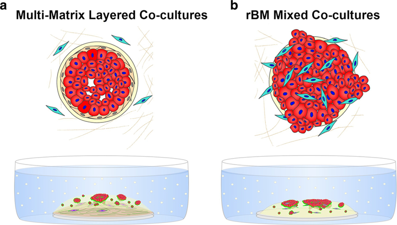
Fig. 1. 3D co-culture models for analysis of DCIS transition to invasive phenotype.

Top: a Architecture of pre-invasive DCIS in which the basement membrane (cream) and layer of myoepithelial cells (MEPs; beige) surrounding DCIS cells (red) are intact. Fibroblasts (cyan) are present in surrounding stroma. b Architecture of microinvasive DCIS in which areas of basement membrane (cream) are disrupted, MEPs (beige) lost, and fibroblasts (cyan) have infiltrated into lesion. Bottom: Pathomimetic 3D co-cultures to model pre-invasive (a) and microinvasive (b) DCIS. a In multi-matrix layered co-cultures, DCIS cells and fibroblasts are cultured in relevant matrix: fibroblasts (fuchsia)in lower layer of type I collagen and DCIS cells (red) ± breast myoepithelial cells (unlabeled) in a top layer of rBM as in a rBM overlay culture. To follow proteolysis, quenched fluorescent substrates (dyequenched (DQ)-collagens IV and I) are mixed with rBM and collagen I, respectively. Green represents the fluorescent cleavage products of these substrates. b In the rBM mixed co-cultures, a mixture of DCIS cells ± MEPs and fibroblasts are plated in a rBM overlay culture. To follow proteolysis, quenched fluorescent substrate (DQ-collagen IV) is mixed with rBM. Green represents the fluorescent cleavage products of these substrates
