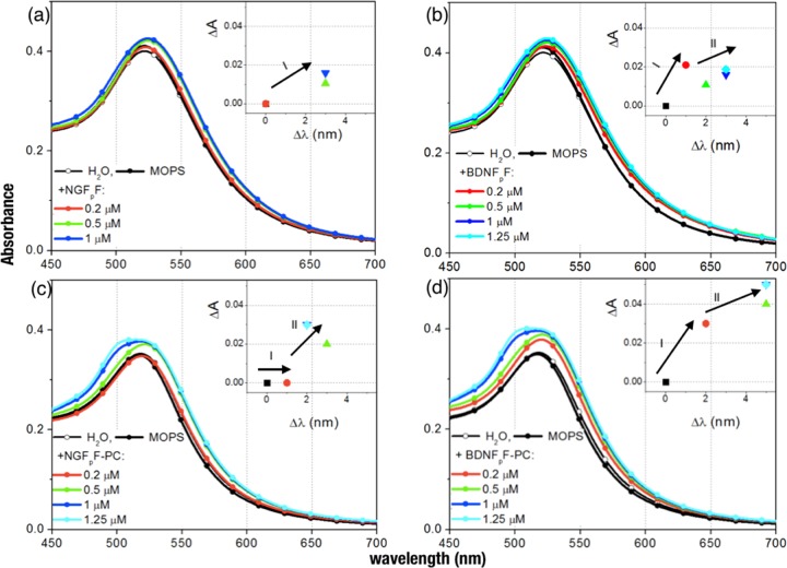
Figure 1.
UV–vis spectra of AuNPs (2.2 nM) before (in water and in 3-(N-morpholino) propanesulfonic acid (MOPS) buffer) and after the addition of peptides at increasing concentrations (range, 2 × 10⁻⁷–1.2 × 10⁻⁶ M) of (a) NGFpF, (b) BDNFpF, (c) NGFpF-PC, and (d) BDNFpF-PC. Insets: ΔA–Δλ scatter plots, with arrows indicating different steps of plasmon shifts (I, II). All experiments are conducted in triplicate.

