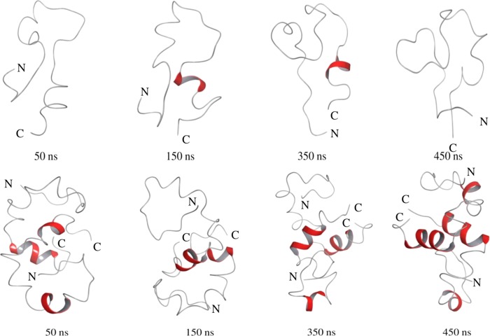
Figure 6.
Snapshots of the conformational state of the Aβ40 peptide monomer (upper panel) and dimer (lower panel) over different time scales of MD simulations in water at 310 K using the Desmond algorithm. The peptide coloring schemes are as follows: red for helical conformation and gray for unstructured region.

