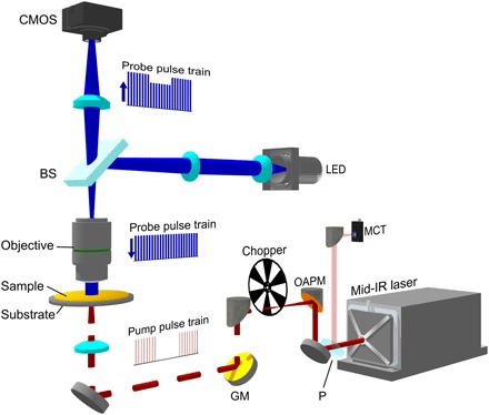
Fig. 1. Schematic of WPS microscope.

A nanosecond mid-IR laser (bottom right) was sent through an optical chopper and weakly focused on the sample. The IR beam was partially sampled with a CaF2 plate (P) and sent to an MCT detector. The probe was provided by a 450-nm LED, which was imaged to the back aperture of an imaging objective by a 4f lens system and a 50/50 beam splitter (BS). The sample-reflected light was collected by the same objective and sent to an image sensor with a tube lens. GM, gold mirror; OAPM, off-axis parabolic mirror; CMOS, complementary metal-oxide semiconductor.
