
Fig. 6. WPS imaging of different chemical components in living cells.
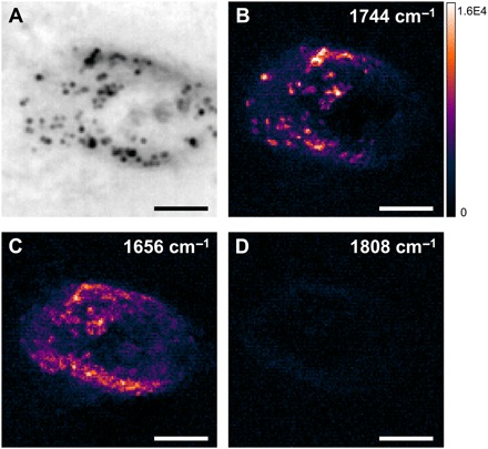
(A) Reflection image of a living SKOV3 human ovarian cancer cell cultured on a silicon wafer. (B to D) WPS images of the same field of view at 1744 cm−1 (lipid), 1656 cm−1 (protein), and 1808 cm−1 (off-resonance), respectively. Scale bars, 10 μm.